Read about Xanthogranulomatous Cholecystitis Ultrasound, MRI, Symptoms, Causes, Treatment.
What is Xanthogranulomatous Cholecystitis?
Xanthogranulomatous cholecystitis (XGC) is an uncommon, persistent inflammatory gallbladder disease. It is characterized by macrophage infiltration and the production of granulomas containing lipid-laden cells known as xanthoma cells. It might be difficult to identify XGC since it can mimic acute cholecystitis clinically and radiologically.
Symptoms
The signs and symptoms of XGC can resemble acute cholecystitis and are typically nonspecific. The following are typical signs:
- Abdominal pain, usually in the right upper quadrant
- Fever
- Vomiting and nausea
- Loss of appetite
- Weight loss
- Jaundice (infrequent)
Causes
The rare inflammatory condition known as Xanthogranulomatous Cholecystitis (XGC) affects the gallbladder. Although the precise cause of XGC is not entirely understood, numerous suggestions have been put forth. According to one hypothesis, bile enters the gallbladder wall as a result of mucosal ulceration or Rokitansky-Aschoff sinus rupture brought on by increased intraluminal pressure as a result of gallbladder or cystic duct obstruction. The rupture of the Rokitansky-Aschoff sinuses and extravasation of bile into the gallbladder is the most commonly accepted scenario for the etiology of XGC.

Diagnostic Imaging
Diagnostic imaging is essential for the assessment of XGC. Ultrasound and magnetic resonance imaging (MRI) are two commonly utilized modalities for assessing the gallbladder and diagnosing XGC.
Ultrasound Imaging
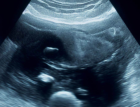
On ultrasound, XGC appears as a thicker gallbladder wall with hypoechoic regions. The gallbladder could seem swollen, nodular, and asymmetrical in form. The presence of intraluminal echoes and movable stones may also be seen. The vascularity of the gallbladder wall can be determined with the aid of color Doppler ultrasound.
MRI Imaging
For determining complications and gauging the severity of XGC, MRI is helpful. On T1-weighted and T2-weighted MRI scans, XGC appears as a thicker gallbladder wall with patches of hypointense or hyperintense tissue. Gallstones, collections of pericholecystic fluid, and involvement of nearby organs can all be seen.
Treatment
XGC is normally treated surgically, while non-surgical treatments may be attempted in some situations.
Surgical Management
The main form of treatment for XGC is cholecystectomy, or surgical removal of the gallbladder. There are typically two major methods employed:
Laparoscopic Cholecystectomy
The preferred surgical method for XGC is laparoscopic cholecystectomy. It entails making small incisions and using a laparoscope to see and remove the gallbladder. Laparoscopic surgery has a number of benefits, including fast recovery, less post-operative discomfort, and shorter hospital stays.
Transverse Cholecystectomy
In circumstances where laparoscopic surgery is technically difficult or inappropriate, an open cholecystectomy may be required. It entails a wider incision with the gallbladder being removed directly. A longer hospital stay and a higher risk of complications are linked to open surgery.


0 Comments