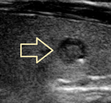
This article is about Punctate Echogenic Foci Thyroid Ultrasound.
A typical diagnostic method for determining the health of the thyroid gland is thyroid ultrasonography. Radiologists may find punctate echogenic foci in the thyroid during these imaging procedures, which might lead them to feel concerned about possible abnormalities.
What Are Punctate Echogenic Foci Ultrasound?
Punctate echogenic foci are small, well-defined, and highly reflecting areas that can be seen on ultrasound scans of the thyroid gland. These foci, which are frequently smaller than 1 mm in size, are distinguished by their bright appearance in contrast to the thyroid tissue around them. Although they can occur in various areas of the thyroid, they are most commonly found in the parenchyma, which is the gland's functional tissue.

Causes
Understanding the etiology of punctate echogenic foci in thyroid ultrasonography is critical for correct diagnosis. Several typical causes and connections are as follows:
Calcifications
Calcifications in the thyroid tissue are frequently the cause of punctate echogenic foci. Benign diseases like thyroid cysts or colloid nodules can cause calcifications. They may, however, occasionally be a sign of serious diseases like papillary thyroid carcinoma or other thyroid cancers.
Colloid Nodules
Non-cancerous growths called 'colloid nodules' may develop in the thyroid gland. These nodules are frequently packed with colloid, a sticky material that aids in hormone synthesis. Punctate echogenic foci may occasionally be associated with the presence of colloid nodules, particularly if the colloid content is high.
Microcalcifications
Small calcium deposits known as microcalcifications can also play a role in the development of punctate echogenic foci. Particularly in papillary thyroid carcinoma, microcalcifications are frequently linked to thyroid cancer.
Significance
In solid and primarily solid nodules, 77.8% of punctate echogenic foci with comet-tail artifacts are malignant. Punctate echogenic foci, as well as clumped calcifications, peripheral calcium deposits, and those with small comet-tail anomalies, are linked to a high incidence of malignancy. They are also among the thyroid cancer indicators.
Evaluation
To ascertain the clinical relevance of punctate echogenic foci found during a thyroid ultrasound, additional investigation is required. Here are some important things to think about:
Fine-Needle Aspiration (FNA) Biopsy
An FNA biopsy may be advised if punctate echogenic foci are found and there are additional characteristics that increase suspicion of malignancy, such as irregular margins or microcalcifications. During this process, thyroid tissue samples are taken for laboratory analysis using a thin needle. Whether the foci are cancerous or benign can be determined using the FNA biopsy results.
Follow-up Monitoring
When punctate echogenic foci seem benign and don't show any alarming signs, diligent observation with frequent ultrasound follow-ups may be advised. This method enables medical practitioners to monitor any alterations in the foci's characteristics or size over time.
Expert Interpretation
Due to the potential diagnostic complications offered by punctate echogenic foci, it is critical to have ultrasound images reviewed by expert radiologists or endocrinologists. Their competence ensures accurate assessments and appropriate management approaches for patients.


0 Comments