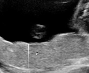
What is Succenturiate Lobe Placenta?
Succenturiate lobe placenta, also known as accessory lobe placenta, is a condition that occurs during pregnancy where there is an additional, smaller lobe of placental tissue separate from the main placenta. Normally, the placenta is a disc-shaped organ attached to the uterine wall that provides oxygen, nutrients, and waste removal for the developing fetus. However, in cases of succenturiate lobe placenta, there is an extra lobe or lobes connected to the main placenta by blood vessels.
The exact cause of succenturiate lobe placenta is not known, but it is believed to occur due to an abnormal division of the placental tissue during early embryonic development. It is relatively rare, with an estimated incidence of about 3-5% of all pregnancies.
Succenturiate Lobe Placenta Ultrasound
During an ultrasound examination, succenturiate lobe placenta can be visualized and diagnosed. Ultrasound is a common imaging technique used during pregnancy to monitor the development of the fetus and assess the health of the placenta.
When performing an ultrasound to detect succenturiate lobe placenta, the sonographer or radiologist will typically look for the following characteristics:
Additional Lobes: They will carefully examine the placenta to identify any additional lobes separate from the main placenta. These additional lobes may be smaller in size and connected to the main placenta by blood vessels.
Vascular Connections: The ultrasound can show the blood vessels connecting the additional lobes to the main placenta. These connections may appear as branching vessels that extend from the main placenta to the accessory lobe.
Location: The ultrasound can help determine the location of the succenturiate lobe within the uterus. It may be located in a different area of the uterus compared to the main placenta.
Blood Flow: The ultrasound may also evaluate the blood flow within the placenta and its lobes. Doppler ultrasound, a specialized technique, can be used to assess the direction and velocity of blood flow in the vessels.

Succenturiate Lobe Placenta Complications
Succenturiate lobe placenta can give rise to several complications during pregnancy and childbirth. Although not all cases result in significant issues, it is important to understand and manage the potential risks. One of the main concerns is the possibility of retained placenta, where the additional lobes remain attached to the uterine wall after delivering the main placenta. Retained placental tissue can lead to complications such as postpartum hemorrhage and infection if not addressed promptly. Another complication associated with succenturiate lobe placenta is placental abruption, where the placenta detaches from the uterine wall before the baby's birth. This condition can cause severe bleeding, endangering both the mother and the baby.
Succenturiate Lobe Placenta Hemorrhage Risk
Succenturiate lobe placenta carries an increased risk of hemorrhage, primarily when the additional lobes are not fully expelled following the delivery of the main placenta. Retained placental tissue is a key factor contributing to this risk. If the additional lobes are not completely delivered, they can result in postpartum hemorrhage, which involves excessive bleeding after childbirth. The abnormal vascular connections between the main placenta and the additional lobes can be prone to bleeding as well, further increasing the risk.


0 Comments