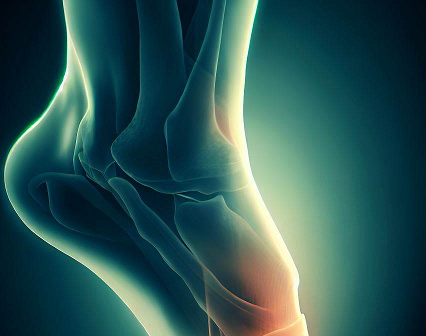
Read about Calcaneofibular Ligament Tear, Sprain, Pain, MRI.
The human body is a masterpiece of complicated structure, with several complex parts that coordinate to facilitate movement and maintain equilibrium. The calcaneofibular ligament, a vital part of the ankle joint, is one such structure.
What is Calcaneofibular Ligament?
The calcaneofibular ligament is a band of connective tissue found on the lateral side of the ankle joint. It is part of the lateral collateral ligament complex, together with the anterior and posterior talofibular ligaments. These ligaments stabilize the ankle and keep the foot from excessively inverting (rolling inward).
Calcaneofibular Ligament Tear
Calcaneofibular ligament tears are often caused by a rapid and abrupt inversion of the ankle. Some of the most common causes are:
- Ankle sprains brought on by sports-related activities or accidents
- Awkward landing after a high jump or fall
- Ankle twisting while moving quickly over uneven terrain
- Ankle joint trauma or impact

Calcaneofibular Ligament Sprain
The term "sprain" describes the stretching or tearing of the calcaneofibular ligament. This injury often happens when the ankle rolls inward forcefully or beyond its normal range of motion, causing undue stress on the ligament. It is a common sports-related injury observed in activities such as basketball, soccer, and jogging.
Calcaneofibular ligament sprain may be caused by a number of factors, such as:
- Running or jumping with abrupt changes in direction
- Landing on an unlevel surface
- Twisting the ankle firmly
- Poor footwear or insufficient ankle support
- weak ankle muscles or past ankle injuries
Calcaneofibular Ligament Pain
Pain in the calcaneofibular ligament can be caused by a number of things. An ankle sprain is the most frequent cause and can be brought on by sports, accidents, or even just stepping on an uneven surface. The ankle joint may experience pain and instability if the ligament is overstretched or damaged. Repetitive strain, overuse injuries, or certain medical disorders that weaken the ligament may also be contributing factors.
Depending on the extent of the injury, the signs and symptoms of calcaneofibular ligament pain may vary. Common symptoms include discomfort on the outside of the ankle, swelling, soreness, bruising, and difficulty bearing weight on the affected foot.
Calcaneofibular Ligament MRI
Several approaches can be used in MRI imaging of calcaneofibular ligament injuries to achieve precise and valuable results. These consist of:
T1-weighted Imaging
Bone and soft tissue features can be assessed using T1-weighted images, which offer great anatomical details. They can be used to spot bone fractures or other abnormalities linked to ligament injury.
T2-weighted Imaging
T2-weighted imaging emphasize fluid-filled structures and aid in the visualization of soft tissue edema, both of which are frequently present in ligament injuries. These images are useful for evaluating ligament damage and other related disorders.
Fat-Suppressed Sequences
To reduce the signal from fatty tissues and make ligament damage more visible, fat-suppressed sequences are utilized. They are especially useful for finding minute tears or undetectable anomalies in the calcaneofibular ligament.


0 Comments